Fig. 3

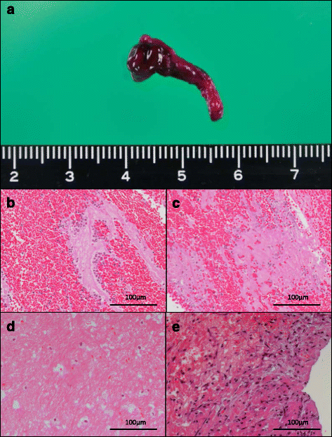
Representative macro- and microscopic images of aspirated deep vein thrombi. a. Representative macroscopic image of an aspirated thrombus. The aspirated thrombus is red or mixed red and white. b. Representative image of a fresh thrombus composed of erythrocyte-rich areas, eosinophilic granular or fibrinous areas, and polymorphonuclear or mononuclear leukocytes. Neutrophils are mainly accumulated at the border of the erythrocyte-rich and eosinophilic areas (9 days after onset). c. Representative image of lytic changes, including the loss of cellular morphology, karyolysis, and karyorrhexis (9 days after onset). d. Representative image of macrophage-like cells (60 days after onset). e. Representative image of an organizing reaction showing fibroblastic/myofibroblastic proliferation, leukocytic infiltration, and matrix deposition (33 days after onset). Hematoxylin and eosin stain (b–d)